By: Yi Zheng, Optometry Intern, Class of 2016
Traumatic brain injuries (TBIs) such as concussions, strokes and whiplash injuries frequently result in oculomotor dysfunction and visual disturbances. Common vision disorders associated with TBIs include deficits in accommodation, version, vergence, visual field, and photosensitivity.1 The intimate interactions between these visual systems can produce symptoms that significantly affect the patient’s ability to function in the workplace and at home. Effective treatment methods are discussed in this article.
Post-TBI patients will often have difficulty functioning in environments with rich visual stimulus; this is sometimes referred to as “supermarket syndrome.” The use of bi-nasal occlusion often works well to alleviate symptoms of visual discomfort, imbalance and vertigo. This treatment method restricts the field of overlapping vision shared between the eyes and reduces visual confusion. A small vertical strip of semi-transparent tape or Bangerter foil is applied to the nasal side of both spectacle lenses. The width of the occlusion is determined by maximizing comfort and mobility.3 Electively, a Streff Wedge can be used to trial the occlusion and for accurate measurement the occluder width.
Visual field inattention is also common in patients that have suffered a TBI. These patients experience symptoms such as bumping into objects or missing words on the affected side when reading.1 Base in prisms have been used to treat these visual field deficits with good success. A 2-3^ BI of ground-in prism is incorporated into the patient’s spectacles. By increasing the divergence demand, the visual system is encouraged to become more aware of the peripheral field of vision.
Padula et al. propose that the visual dysfunction caused by TBIs is caused by impaired ambient (magnocellular) processing. Their study showed that treatment with bi-nasal occlusion and base in prism increases the amplitude on VEP (P100) for post-TBI patients, which suggests improved function in the binocular cortical cells. In addition, the symptoms improved for patients subjectively.2
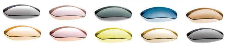
Further, solid tints are useful to help relieve photosensitivity that some patients experience. Colours are selected by subjective trialing. Popular choices include brown, grey, amber and blue.1 The above treatment options have had lots of success in the management of post-TBI associated visual dysfunction. They can be easily incorporated into eye exams and trialed.
Finally, vision therapy is an excellent option to explore to address the underlying problem. A customized vision therapy program can work to mend accommodative, oculomotor, visual field (scanning therapy), and visual processing dysfunction at a patient-specific pace and minimize the rehabilitation process after a TBI. For more information, go to www.covd.org
References
- Kapoor, Neera, and Kenneth J. Ciuffreda. "Vision disturbances following traumatic brain injury." Current
Treatment Options in Neurology 4.4 (2002): 271-280.
- Padula, William V., S. Argyris, and J. Ray. "Visual evoked potentials (VEP) evaluating treatment for
Post-trauma vision syndrome (PTVS) in patients with traumatic brain injuries (TBI)." Brain Injury 8.2 (1994): 125-133.
- Proctor, Alissa. "Traumatic Brain Injury and Binasal Occlusion." Optometry & Vision Development40.1
